Definition:
Otitis externa (OE) is an intense inflammation of the External auditory Canal
Microorganisms
- Most common microorganism: Pseudomonas aeruginosa
- Other common organisms: Staphylococcus aureus and Staphylococcus epidermidis
- Uncommon microorganisms: Coryneform (diphtheroids), Gram-negative rods (Enterobacter Klebsiella, Proteus, Escherichia coli), Streptococcus and Enterococcus.
Actinomyces israelii is an anaerobic Gram-positive bacterium that can cause OE from primary dental or parotid infection.
Refractory OE presents with granulation tissue in EAC and thick yellow ear discharge and needs surgical debridement and prolonged antibiotic therapy.
Cultures for bacteria and fungus are indicated in persistent or refractory infection.
Acute Otitis Externa
Excessive and repeated removal of the hydrophobic earwax coating of EAC exposes the underlying epithelium to water, infection, and other contaminants and leads to progressive erythema and edema.
Predisposing factors
1) Warm humid environment:
Excessive sweating changes the canal pH to alkaline, which favors bacterial growth.

2) Obstruction of EAC:
Stenosis, exostoses, impacted wax are major contributing factors for OE.
3) Ear trauma:
Use of cotton-tip swabs, irrigators for wax removal, in-the-canal hearing aids, digital ear thermometers, unskilled instrumentation.
4) Water contamination of EAC:
It is usually swimming related.

Hence, acute otitis externa is also very commonly known as the SWIMMER’s EAR.
Clinical features
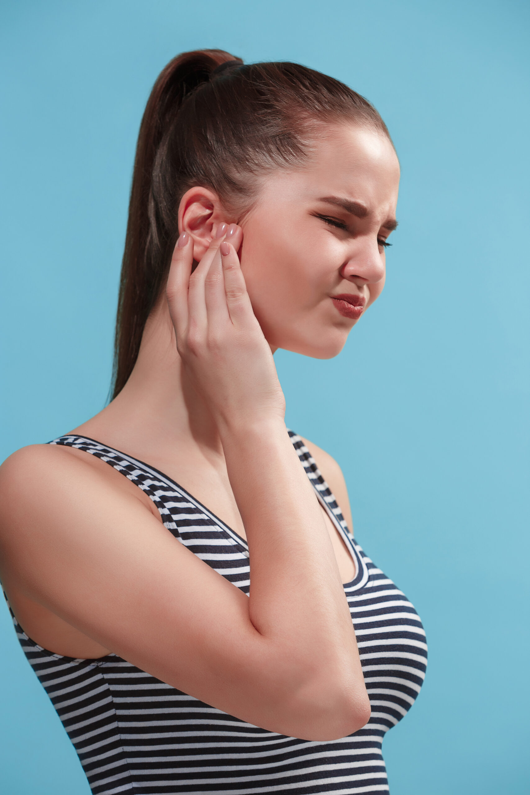
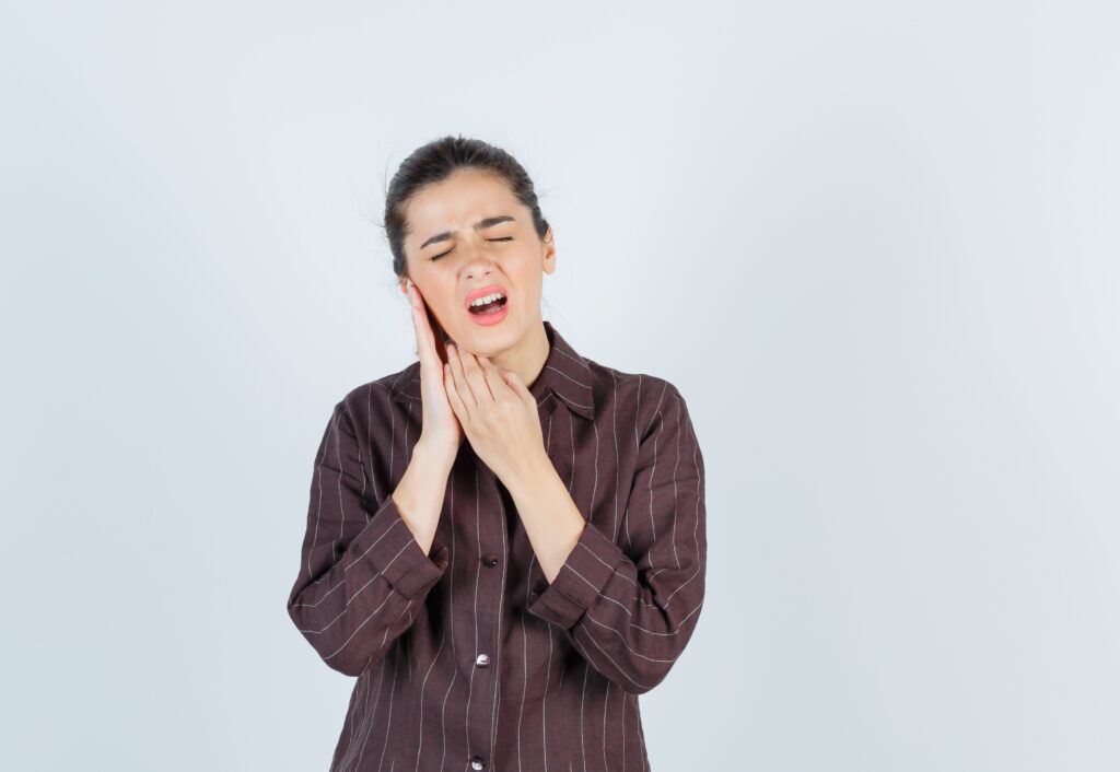
1) INTENSE AND SEVERE EAR PAIN
It is the chief presenting complaint.
Ear pain may be aggravated with jaw movements like chewing, opening the mouth, etc.
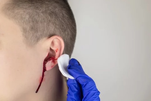
2) EAR DISCHARGE
It is usually watery-like means serous.
3) Itching
It is present in some cases.
STAGES OF OTITIS EXTERNA
Early stage: An erythematous canal with scant discharge
Later on: Edematous and exquisitely tender canal occluded with purulent squamous debris
In severe cases: Enlarged and tender regional lymph nodes and periauricular cellulitis
No additional systemic sign and symptoms
Treatment of Otitis Externa
1) Cleaning of EAC
This clears the infected debris material and allows topical antibiotics to reach the infected site.
2) Acidification of EAC
Acetic acid solution and antibiotics in acidic suspensions and solutions kills many bacteria and fungus.
3) Topical Antibiotics
Topical Antibiotics ear drops provide a concentration many times greater than systemic antibiotics in the ear canal. Thus usually it is not necessary to start systemic antibiotics even in resistant infection.
Quinolone antibiotic drops (ofloxacin, ciprofloxacin) with dexamethasone (hydrocortisone is avoided as it leaves a precipitate) in an acidic vehicle are the best as they have no ototoxicity and no potential for allergic reactions.
Antibiotic impregnated ear wicks (cotton, merocel, ribbon gauze) offer better absorption through an edematous canal skin.
4) Oral antibiotics
These are reserved for complications of OE such as cellulitis, perichondritis, chondritis and malignant OE.
Chronic Otitis Externa
No prior history of trauma or water contamination
Usually not painful
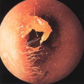
Examination may reveal FUNGAL HYPHAE in the canal or keratin debris from chronic dermatitis.
Acute exacerbation is usually preceded by itching.
Types of Chronic OE
The four different disease states with distinct etiologies are allergic, contact dermatitis, seborrhea and granular OE.
1) Allergic OE
Clinical features:
- Edema and purulent discharge secondary to topical antibiotic (most commonly neomycin) ointment or drops.
- Maculopapular eruption in concha and EAC.
- A thickened and erythematous canal.
Treatment of Allergic OE:
– Elimination of offending agent, debridement and topical steroid drops.
2) Contact Dermatitis
Clinical features:
- It can result from a variety of agents, such as hairsprays, shampoos and hearing aid molds.
- A thickened and erythematous canal.
Treatment of Contact Dermatitis:
–Elimination of offending agent.
– Debridement and topical steroid drops.
3. Psoriasis or Seborrhea
Clinical features:
- Hyperkeratosis and lichenification of EAC skin.
- Seborrheic OE is associated with seborrheic dermatitis of the scalp.
- Itching is the presenting complaint.
- Greasy yellow scales may be seen in the canal, over the lobule, and in the postauricular sulcus.
- Secondary bacterial or fungal infections may occur.
Treatment of Seborrheic OE:
– Treatment of scalp seborrhea.
– Ear toilet, salicylic acid, and sulphur cream
4. Granular OE
Clinical features:
- It results from chronic bacterial or fungal infection of EAC.
- Granulation and excoriation seen on TM and EAC skin.
- Culture for bacteria and fungus may show causative organisms.
Treatment of Granular OE-
– Repeated debridement.
– Cauterization of granulations.
– Topical antibiotic or antifungal creams.
– Topical gentian violet facilitates drying the EAC.
– Topical steroid or 5-fluorouracil.
– De-epithelialization of TM: If does not respond to drying agent (Burow’s solution) it may need spilt thickness skin graft.
Complications of OTITIS EXTERNA
1) Cellulitis:
It presents as an erythematous ear. There is no induration like perichondritis.
Treatment is with systemic antistaphylococcal agents.
2) Perichondritis or Chondritis
3) Medial canal fibrosis:
In chronic otitis externa (COE), a thick fibrous scar may obstruct the deep aspect of EAC.
TM appears lateralized with the absence of typical landmarks.
Surgical treatment consists of excision of the fibrous scar followed by canaloplasty with split-thickness skin graft and if needed, tympanoplasty.
4) Malignant otitis externa
A very severe LIFE THREATENING form of Otitis Externa.
THANK YOU
MEDICAL ADVICE DISCLAIMER:
This blog, including information, content, references, and opinions, is for informational purposes only.
The Author does not provide any medical advice on this platform.
Viewing, accessing, or reading this blog does not establish any doctor-patient relationship.
The information provided in this blog does not replace the services and opinions of a qualified medical professional who examines you and then prescribes medicines.
If you have any questions of a medical nature, please refer to your doctor or qualified medical personnel for evaluation and management at a clinic/hospital near you.
The content provided in this blog represents the Author’s own interpretation of research articles.